
Home / Reporting
New to the app
Three studies, entered in full
These are finished screens, not empty forms. After you sign in, open the matching tool, type what you measured, and read the suggestion before it goes into the report.
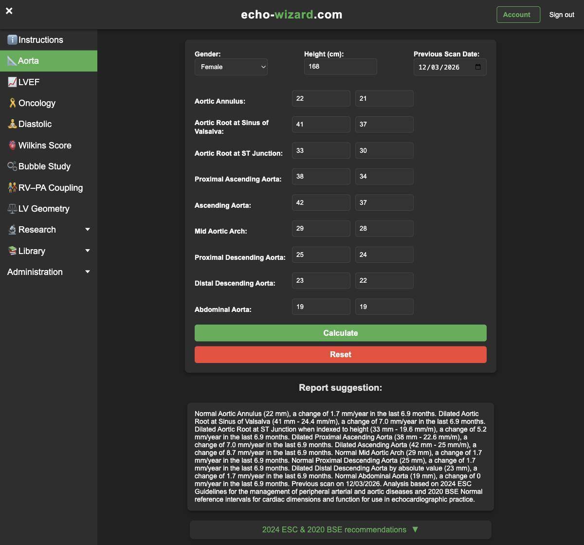
Aorta, every segment, with the previous study
Sex, height and the date of the last scan sit at the top. Each segment has a current measurement and the previous one. Leave a box empty only when you did not measure it. The wording draws on the 2024 ESC aortic guideline intervals and the 2020 BSE reference intervals. That is a source for the cut-off, not an endorsement of this app.
In this example the patient is female, height 168 cm, previous scan 12 March 2026. Annulus 22 mm (was 21). Sinus of Valsalva 41 mm (was 37). Sinotubular junction 33 mm (was 30). Proximal ascending aorta 38 mm (was 34). Ascending aorta 42 mm (was 37). Mid arch 29 mm (was 28). Proximal descending 25 mm (was 24). Distal descending 23 mm (was 22). Abdominal aorta 19 mm, unchanged.
The suggestion begins: “Normal Aortic Annulus (22 mm), a change of 1.7 mm/year in the last 6.9 months. Dilated Aortic Root at Sinus of Valsalva (41 mm - 24.4 mm/m), a change of 7.0 mm/year in the last 6.9 months.” It continues through the ascending aorta at 42 mm (25 mm/m), the arch, both descending segments and the abdominal aorta, and it names the previous scan date.
Cardio-oncology, four scans and both ventricles
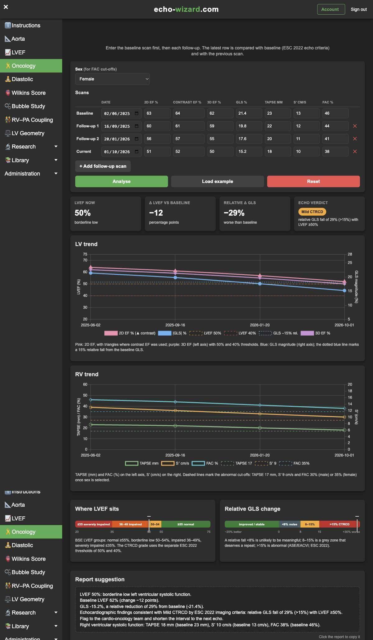
Enter the baseline first, then each follow-up. Every row can hold the date, 2D ejection fraction, contrast ejection fraction, 3D ejection fraction, GLS, TAPSE, S′ and fractional area change. The latest row is compared with baseline. The charts use the whole series.
This series is female. Baseline 2 June 2025: 2D EF 63%, contrast 64%, 3D 62%, GLS 21.4%, TAPSE 23 mm, S′ 13 cm/s, FAC 46%. Follow-up 16 September 2025: 60, 61, 59, 19.8, 22, 12, 44. Follow-up 20 January 2026: 56, 57, 55, 17.6, 20, 11, 41. Current 1 October 2026: 51, 52, 50, 15.2, 18, 10, 38.
Because 3D ejection fraction is present at both ends, the suggestion uses it: “LVEF 50%: borderline low left ventricular systolic function. Baseline LVEF 62% (change −12 points). GLS -15.2%, a relative reduction of 29% from baseline (-21.4%). Echocardiographic findings consistent with Mild CTRCD by ESC 2022 imaging criteria: relative GLS fall of 29% (>15%) with LVEF ≥50%.” The right ventricle is in the same paragraph: TAPSE 18 mm from 23, S′ 10 cm/s from 13, FAC 38% from 46%.
Bubble study, from the septum to the grade
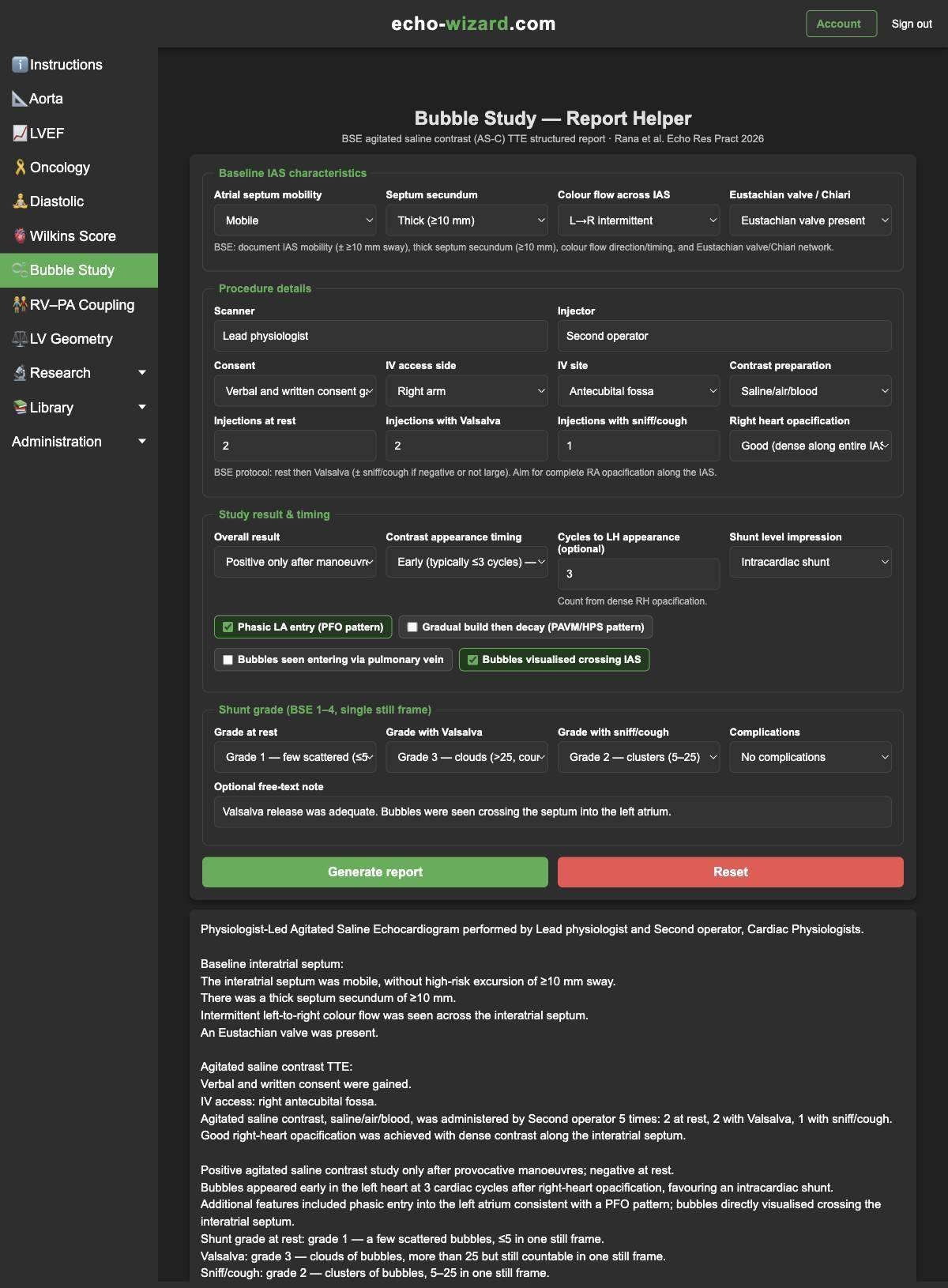
The bubble tool follows a structured agitated-saline report: baseline septum, who scanned and who injected, consent, access, how many injections, opacification, timing, shunt level, the extra signs, and the grade at rest, with Valsalva and with sniff.
In this example the septum is mobile, the septum secundum is thick (≥10 mm), colour flow is intermittent left to right, and an Eustachian valve is present. Consent is verbal and written. Access is the right antecubital fossa. Contrast is saline, air and blood: 2 injections at rest, 2 with Valsalva, 1 with sniff. Right-heart opacification is good. The result is positive only after a manoeuvre, appearance is early, at 3 cycles, and the impression is an intracardiac shunt. Phasic left-atrial entry and bubbles crossing the septum are ticked. Grade 1 at rest, grade 3 with Valsalva, grade 2 with sniff. No complications.
The suggestion opens “Physiologist-Led Agitated Saline Echocardiogram” and ends: “Positive agitated saline contrast study only after provocative manoeuvres; negative at rest. Bubbles appeared early in the left heart at 3 cardiac cycles after right-heart opacification, favouring an intracardiac shunt.” The three grades are spelled out underneath.
Which tool for which part of the report
| What you are writing | Open this |
|---|---|
| Aortic annulus through the abdominal segment | Aorta. Current and previous measurements, sex and height. |
| Serial ejection fraction and GLS on cancer therapy | Cardio-oncology. One row per scan, including contrast, 3D and the right ventricle. |
| Agitated-saline shunt study | Bubble study. Septum, injections, timing, grade and the overall result. |
| Left-ventricular size and wall thickness | LV geometry. Sex, height, weight, LVIDd, IVSd and PWd. |
| Ejection fraction, GLS, TAPSE, S′, FAC | Ventricular function, with baseline and previous values when you have them. |
| Mitral valve morphology before balloon valvuloplasty | Wilkins score. Leaflet mobility, thickening, calcification and the subvalvular apparatus. |
| Diastolic function, one question at a time | Diastolic assistant. The pathway stays on the server. |
| Diastole when the mitral valve is diseased | Diastolic index, using IVRT and TE-e′. |
| TAPSE against pulmonary pressure | RV–PA coupling, with the cut-off you choose. |
What you still do yourself
Look at the images. Decide whether the suggestion is the line you would sign. Research screens — myocardial work, HFpEF scores, TEER measurements, amyloid pattern, and the rest — structure a discussion. They are not the clinical conclusion, and several of them say so on the page.
Reference posters for chambers, valves, diastole and amyloid are available while a membership is active. They sit beside the report. They are not a substitute for the guideline you follow locally.
For informative use only. Echo Wizard is for fully trained echocardiographers. You remain responsible for checking every number before it goes into a report.