1. Open the matching tool
Aorta, cardio-oncology, bubble study, prosthetic valve, geometry, ventricular function, diastole, Wilkins, RV–PA coupling. Research tools sit in their own group.
 echo-wizard.com
Echocardiography reporting
Type in what you measured on the study you are reporting, and the previous study when the comparison matters. Echo Wizard drafts the paragraph for that part of the report. You check it against the images and copy it across.
No card needed to start. Built for echocardiographers, cardiac physiologists, clinical scientists and cardiologists. The report stays yours.
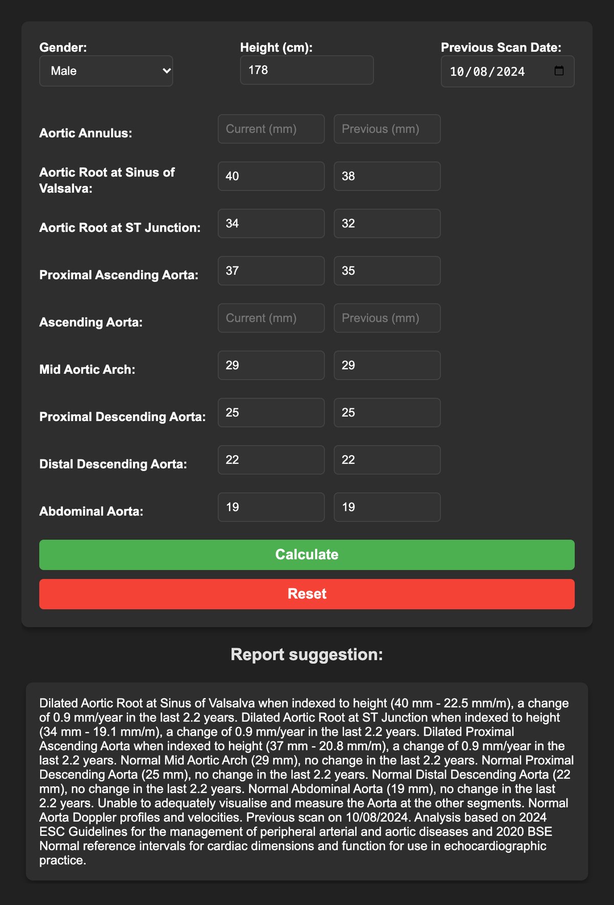
Aorta. Bicuspid aortic valve surveillance, 2.2 years on: the root and proximal ascending aorta have grown a little, the rest has not changed.
The slow part is rarely the measuring. It is the comparison, the indexed value, and the paragraph you would otherwise assemble from memory between patients.
Enter sex, height, and each segment you measured today and last time. The suggestion gives the absolute size, the indexed size, and the change in millimetres per year. In the example above, a man with a bicuspid valve, the sinus of Valsalva is 40 mm (22.5 mm/m), up from 38 mm 2.2 years earlier: 0.9 mm/year. The arch and descending aorta read “no change”.
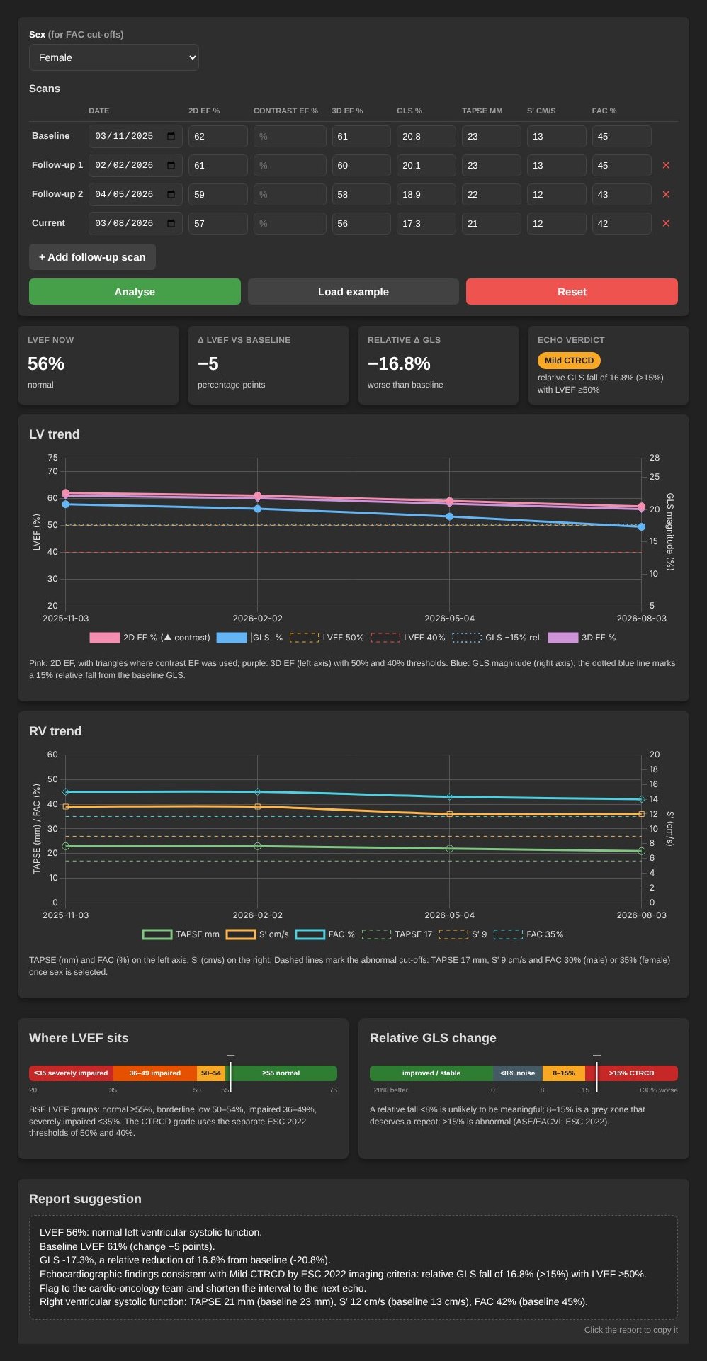
One row per scan: date, 2D ejection fraction, contrast, 3D, GLS, TAPSE, S′ and fractional area change. The latest row is set against baseline. The example is a woman on trastuzumab scanned every three months: LVEF stays above 50%, but GLS falls by 16.8% from baseline, which meets the ESC 2022 definition of mild CTRCD.
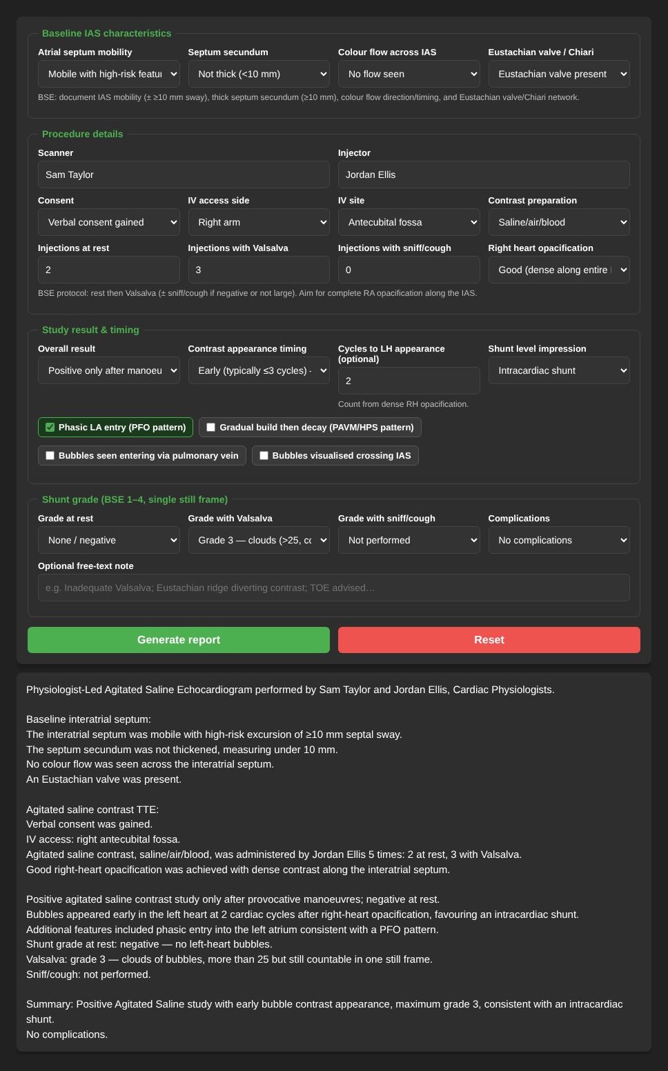
The form follows the newest BSE agitated saline guidance (Rana et al., Echo Res Pract 2026), so every report covers the septum, consent, injections, timing, shunt level and the grade in the same order. The example is a stroke work-up: negative at rest, early grade 3 with Valsalva, consistent with an intracardiac shunt.
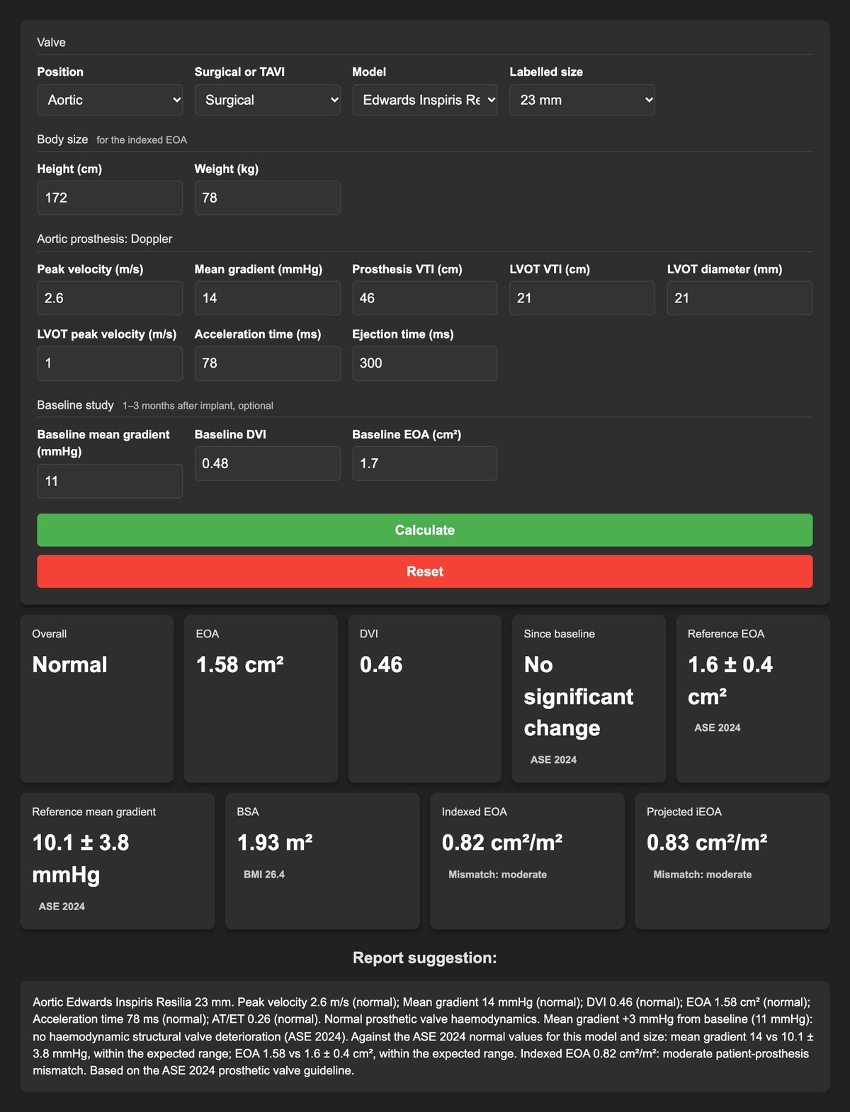
Aortic, surgical or TAVI, mitral, pulmonary and tricuspid, each graded against the ASE 2024 criteria for that position. Pick the model and size and the measurements are set against its published normal values: 121 models, 502 sizes. The example is a 23 mm Inspiris Resilia a year after surgery: normal function, no change from baseline, but an indexed EOA of 0.82 cm²/m², moderate patient-prosthesis mismatch.
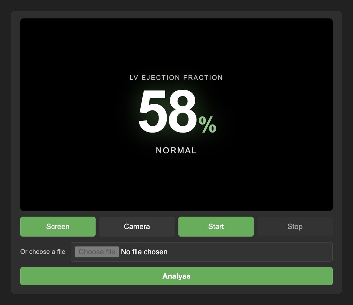
Share the apical 4-chamber loop from your reporting software, frame the left ventricle, and a deep-learning video model returns an EF estimate and its category. It is included with an active membership, free trial or paid, and it sits beside your own measurement rather than replacing it.
Strain, congestion, device measurements and infiltrative patterns, built from recently published research for the unusual case. They are labelled as research and never presented as the clinical report.
Aorta, cardio-oncology, bubble study, prosthetic valve, geometry, ventricular function, diastole, Wilkins, RV–PA coupling. Research tools sit in their own group.
Put in the measurements you acquired, and the earlier ones when the comparison matters. Leave a field blank when you did not measure it.
Read the suggestion against the images. If it matches, copy it. If it does not, change it before it reaches the report.
See five examples entered in full, with the report text each one produces
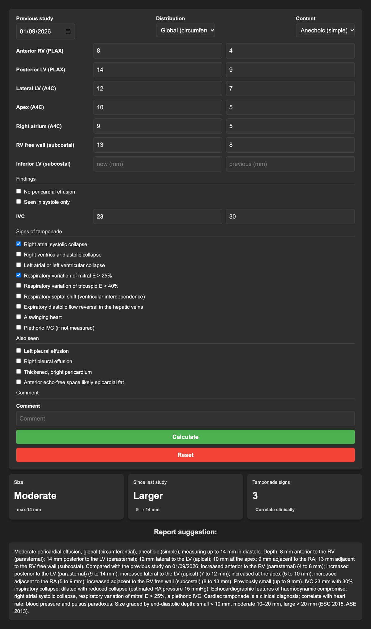
Included with membership
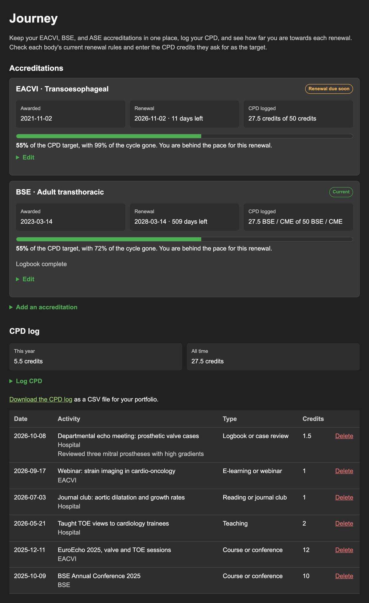
Keep your BSE, EACVI and ASE accreditations together, log CPD as you go, and see how close you are to each renewal.
Renewal rules belong to each body. Enter the CPD target they set, and Journey tracks you against it.
Membership
£29 a year
Every clinical tool, the instructions, the reference posters and Journey for your accreditations and CPD. A new account starts with 7 days free, and no card is needed to start.
Invite a colleague with your referral code: they start with 30 days free, and you get 30 days added once they confirm their email.
You enter the measurements from the study. It drafts the report text for that part of the examination. You read it against the images, change anything that does not match, and copy it into the report.
Aortic size by segment, with the previous study and the change per year. Cardio-oncology, with 2D, contrast and 3D ejection fraction, GLS and right-ventricular measures on every visit. Agitated-saline bubble studies, from the septum through to the shunt grade. Also left-ventricular geometry, ventricular function, the Wilkins score, diastolic function and RV–PA coupling. Research screens are separate and labelled as research. Auto EF, currently in testing, estimates LVEF from an apical 4-chamber clip.
No. It is for fully trained echocardiographers, cardiac physiologists, clinical scientists and cardiologists. It drafts wording from the numbers you enter. You remain responsible for every number and every line in the report.
The clinical tools send what you type to the server, draft the text, and do not keep that calculation as a patient record. Echo Wizard is not the archive of the study.
Fully trained echocardiographers, cardiac physiologists, clinical scientists and cardiologists reporting transthoracic studies. Membership is £29 a year. A new account starts with 7 days free, and no card is needed to start.
Yes. Every member has a referral code on their account page. A colleague who signs up with it starts with 30 days instead of 7, and you get 30 days added once they confirm their email, up to six times a year.
For informative use only. Echo Wizard is for fully trained echocardiographers, cardiac physiologists, clinical scientists and cardiologists. It drafts report text from the measurements you enter so a department can keep its wording consistent. It is provided as is. You remain responsible for checking every number before it goes into a report, and it does not replace your own judgement.